Thursday, 26 June 2025

Story of Vanderwaal forces and its help in coagulation.


πŸ§ͺ “The Tale of the Two Tiny Travelers”

In the magical world of Colloidia, countless tiny particles—called Colloids—float happily in water. Two such particles, Rishi and Meera, were curious adventurers. They had never met, but one day, they started drifting toward each other in the watery world.

⚡️Obstacle #1: The Invisible Wall (Electrostatic Repulsion)

As Rishi and Meera got closer, they suddenly felt a strong invisible push—like two magnets trying to touch but being forced apart.

“What’s happening?” Rishi asked, sliding backward.
“It’s our electric coats!” said Meera. “We’re both wearing negative charges—they don’t like each other!”

Their electric coats were part of their electrical double layer, a protective force field that kept them apart. The closer they tried to get, the stronger the push became. It was like trying to climb a hill that kept growing steeper—the energy barrier!

πŸ’₯The Leap of Faith (Overcoming the Barrier)

But then, a sudden current of water rushed in—a collision! Rishi and Meera were pushed together with such force that they jumped over the invisible wall.

“Hold on tight!” yelled Meera as they flew past the peak.

This strong push gave them enough kinetic energy to overcome the repulsive force. Once past that point, something unexpected happened...

πŸ’žStuck Together Forever (van der Waals Attraction)

As soon as they got really close, they felt a cozy pull—like a warm hug.

“Woah! What is this gentle tug?” asked Rishi.
“It’s the van der Waals force,” Meera whispered. “It’s weak alone, but strong when many molecules pull together.”

Now that they were close enough, there were no more repulsions, only a sticky attraction that trapped them together—an energy trap. They couldn’t escape.

“I guess we’re stuck,” smiled Rishi.
“Forever flocculated,” laughed Meera.

And so, Rishi and Meera became part of a growing floc, a family of particles bound by invisible forces—living proof of the balance between repulsion and attraction in the kingdom of Colloidia.

🌟 Moral of the Story:

Colloidal particles stay apart because of repulsive forces, but if they overcome the energy barrier, attractive forces trap them together. This balance of forces is what DLVO Theory is all about.

Sunday, 8 June 2025

Meera and the Five DNA Processors: A Journey Through the Polymerase City


Once upon a time in the DNA Repair City, lived a curious young scientist named Meera. This city was home to a long and precious scroll called DNA, which carried all the instructions needed to run the city. But this scroll was very delicate and often got little tears, smudges, or missing letters. Luckily, five superheroes known as Polymerase Protectors (Pol I to V) guarded the DNA scroll.

πŸ§ͺ Chapter 1: Pol I – The Cleanup Master (Gene: polA)

One day, Meera saw a superhero with a magnifying glass and scissors, carefully removing little sticky notes (RNA primers) from the scroll and replacing them with proper DNA. This was Pol I, the Cleanup Master.

“I’m not fast,” Pol I told Meera, “but I’m good at cleaning up mistakes. I can also fix small nicks and fill gaps. See? I even proofread!”

Pol I had three tools:

  • A pen to write DNA (5'→3' polymerase),

  • An eraser to remove wrong letters from the end (3'→5' proofreading),

  • And scissors to snip out damaged or RNA parts from the start (5'→3' exonuclease).

Meera noted down: Gene: polA — For primer removal and DNA repair.

πŸ”¬ Chapter 2: Pol II – The Quiet Rescuer (Gene: polB)

Next, Meera met a calm and steady hero, Pol II, hiding in the background.

“I come when DNA is hurt or stuck,” said Pol II. “When the main worker gets stuck, I step in and gently fix things.”

Pol II specialized in restart repair and careful copying after stress. He wasn't the main worker, but always ready in emergencies.

Meera’s notes: Gene: polB — For DNA repair and stress recovery.

⚙️ Chapter 3: Pol III – The Speed Star (Gene: dnaE and friends)

Suddenly, a blur zoomed by! Meera saw a high-speed hero with multiple arms — Pol III, the Main Builder of the city.

“I’m the fastest! I build most of the DNA scroll during replication,” said Pol III proudly. “But I don’t work alone. I have a whole team!”

Pol III had:

  • A pen hand (alpha, dnaE) to write,

  • A proofreading hand (epsilon, dnaQ) to catch errors,

  • A steadying hand (theta, holE) for support,

  • And a sliding clamp that helped him stay attached like Velcro!

Meera gasped: “You’re the main construction crew!”

Gene: dnaE, dnaQ, holE — Main replicative polymerase.

🧫 Chapter 4: Pol IV – The Risk-Taker (Gene: dinB)

Next, Meera heard a loud crash. A daring, slightly clumsy superhero was jumping over broken parts of DNA!

“I’m Pol IV,” he said. “Sometimes when DNA has damage, someone’s gotta keep going. I take risks and fill in the gap — even if it’s not perfect.”

He was part of the SOS response — emergency services when the city is under attack from UV rays or chemicals. He could bypass lesions, but often introduced typos.

Meera’s notes: Gene: dinB — For translesion synthesis (damage tolerance).

⚠️ Chapter 5: Pol V – The Emergency Fighter (Genes: umuC and umuD)

In the deepest part of the city, Meera met the final hero — a serious and slightly dangerous one. It was Pol V, created only in extreme emergencies.

“When DNA is severely damaged and no one else can help,” said Pol V, “I step in. I’m not accurate, but I’ll make sure the scroll is complete, no matter what.”

Pol V worked as a team: UmuD’2C — a combination of two UmuD and one UmuC. Together, they acted like a desperate repair crew under fire.

Meera’s notes: Genes: umuC and umuD — for SOS-induced error-prone DNA synthesis.


🧠 Meera’s Final Summary:

Polymerase Gene(s) Role in DNA City
Pol I polA Removes RNA primers, fills gaps, repairs
Pol II polB Helps in DNA repair and restart
Pol III dnaE, dnaQ, holE Main enzyme for DNA replication
Pol IV dinB Damage bypass during stress (error-prone)
Pol V umuC, umuD Emergency damage bypass (error-prone, SOS)

And with that, Meera closed her notebook, grateful to have met the five polymerase protectors who kept the DNA City safe and functioning. She now understood that each had a special job, and together they made sure life continued smoothly — even when things went wrong.

Meera and the DNA Replication Party: A Story of ORC, Cdc6, Cdt1, and MCM2-7


In the enchanting world of biology, there's a magical event that happens every time a cell prepares to divide — it must make an exact copy of its DNA. But this isn’t just mindless duplication. It’s a highly coordinated royal event, with strict security, special permissions, and a team of molecular workers who make sure everything runs smoothly.

Let me introduce you to Meera, a bright and curious girl who asked a simple question:
"How does a cell know where and how to start copying its DNA?"

Let’s follow Meera’s journey into the microscopic kingdom of Cellandia, where she met the key characters involved in DNA replication licensing.

🏰 Step 1: Meet the Gatekeepers — ORC1 to ORC6

At the gates of the DNA palace stood the Origin Recognition Complex (ORC), a team of six loyal guards: ORC1, ORC2, ORC3, ORC4, ORC5, and ORC6.

Their job?
To identify and bind to the “starting point” on DNA, called the replication origin.

They don’t start the replication themselves. Instead, they mark the spot and say:

“This is the official shelf from where we’ll begin copying the DNA book.”

These ORC proteins stay attached like security guards, making sure the spot is ready when the time is right.

πŸ” Step 2: The Organizer Arrives — Cdc6

Next comes Cdc6, a powerful recruiter who joins hands with ORC.

He carries ATP (cellular energy) and helps build a platform for the next players.
He says:

“Alright team, let’s prepare the origin for the main crew!”

Once Cdc6 does his job — especially during the G1 phase of the cell cycle — he steps back. When the S phase starts, he’s either phosphorylated or removed so he doesn’t cause any trouble. After all, we only want one copy of the DNA, not two or three!

πŸŽ’ Step 3: The Guide — Cdt1

Then comes Cdt1, a gentle guide and a crucial chaperone.

Cdt1’s role is to escort the MCM2-7 helicase — the team that will eventually open up the DNA strands. She gently says:

“MCM friends, follow me. We need to get into position around the DNA.”

She helps load the MCM2–7 hexamer onto the DNA. These are the ring-shaped helicase proteins that form the core of the future replication machinery.

But like Cdc6, Cdt1’s activity must be carefully controlled. That’s where the next character steps in.

🚫 Step 4: The Guardian — Geminin

When the S phase begins, a wise and protective molecule named Geminin shows up.

Her job?
To block Cdt1 from loading more MCM2-7 helicases once replication has started. She sternly reminds the team:

“Only one license per origin. No second chances!”

This ensures the DNA is copied only once per cycle, preserving the cell’s integrity.

πŸ”„ Step 5: The Helicase Heroes — MCM2 to MCM7

Finally, the stars of the show take the stage: MCM2, MCM3, MCM4, MCM5, MCM6, and MCM7.

These six proteins form a ring-shaped helicase complex — the engine that will unzip the DNA strands during replication.

They’re loaded onto DNA in an inactive form during G1, but once S phase begins, they’re activated by other factors (Cdc45 and GINS) to form the mighty CMG helicase, which unwinds DNA so that the polymerases can begin copying it.

πŸŽ‰ Meera’s Takeaway

After meeting all these fascinating molecular players, Meera sat down and wrote in her journal:

“DNA replication is not random. It starts at licensed origins. ORC marks the site, Cdc6 organizes, Cdt1 brings in the helicase, and MCM2–7 unwinds the DNA. Everything happens once per cycle, under strict regulation. It’s like a perfectly managed party — no uninvited guests, no repeated entries.”

She smiled, now understanding how our cells ensure faithful DNA copying, maintaining genome stability and preventing chaos.

πŸ“˜ Final Thoughts

This biological ballet happens in every dividing eukaryotic cell, from humans to yeast. Any mistake in this licensing process can lead to re-replication, mutations, or cancer. That’s why nature has evolved this elegant, tightly controlled system.

So the next time you hear terms like ORC, Cdc6, Cdt1, or MCM2-7, just remember Meera and the DNA replication party — a perfect blend of timing, teamwork, and trust.

Wednesday, 28 May 2025

Meera and the Mystery of DNA Replicon


In a small village filled with sunlight, green fields, and the quiet hum of nature, lived Meera, a bright and curious science student. Her world wasn’t just about books and exams. She saw science everywhere---the buzzing bees made her think of communication, the curling tendrils of vines reminded her of DNA spirals, and the clear night sky made her wonder about life at the microscopic level.

A Wish Under the Banyan Tree

One afternoon, under the cool shade of the village banyan tree, Meera flipped through her biology textbook. She paused at a diagram showing how cells copy their DNA before they divide.

“I wish I could see "How do cells know where and how to start copying their DNA?" she whispered.

Suddenly, the world shimmered. In the blink of an eye, Meera found herself inside a bacterial cell---E. coli.

Inside a Bacterial Cell

In a flash, Meera found herself inside a bacterial cell. All around her were cellular structures like Golgi, Ribosome, ER etc.. she had only seen in diagrams. "She entered into the world of nucleus..."

It was like stepping through the gates of a tightly guarded city, where the blueprint of life was etched into coils of DNA. Surrounded by a nuclear envelope, studded with pores like watchful sentinels, she found herself in the heart of the cell---the command center. 

 A glowing spiral ladder--DNA of E.Coli sparkled softly to surprise Meera.

To her amazement, the DNA began to speak.

The DNA Speaks: I Am a Replicon

“Meera, I am about to replicate. Would you like to watch?”

Meera nodded eagerly.

“I am a replicon,” said the DNA. “That means I am a segment of DNA that contains everything needed to start and finish replication on my own.” I'm also called as unit of replication. 

Bacteria like E. coli have just one replicon that is usually circular. This is known as mono-repliconic.

Eukaryotic cells like humans have many replicons per chromosome, mainly linear. This is known as multi-repliconic, since their DNA is much longer and needs multiple starting points to copy efficiently.

Genome Size Comparison

  • E. coli has approximately 4.6 million base pairs (4.6 x 10⁶ bp).

  • Humans have about 3.2 billion base pairs (3.2 x 10⁹ bp).

Finding the Starting Line: OriC

A small region on the DNA began to glow.

“This is my OriC,” said the DNA. “It’s the origin of chromosomal replication in E. coli. Only 245 base pairs long, but it’s where everything begins bidirectionally or unidirectionally.”

OriC is a cis-acting sequence is a regulatory DNA region that doesn’t move or code for proteins but helps control essential processes on the same DNA molecule.

Inside OriC

Meera saw two kinds of short DNA sequences:

  • Five 9-mer sequences with the code TTATCCACA, known as DnaA boxes. These are the binding sites for DnaA initiator proteins.

  • Three 13-mer sequences rich in adenine and thymine. These are easier to unwind because A-T base pairs have only two hydrogen bonds.

The Initiator Proteins Arrive

DnaA proteins soon arrived, attaching themselves to the 9-mer boxes. This caused the DNA to bend and build up tension, which helped unwind the nearby 13-mer region.

Then came DnaB helicase, which continued unwinding the DNA, paving the way for the full replication machinery.

The Gatekeeper: Methylation at GATC

Suddenly, Meera noticed certain sequences glowing: GATC sites.

“These are special,” said the DNA. “An enzyme called Dam methylase recognizes these sites and adds a methyl group to the adenine base.”

This process is known as methylation at GATC. It acts like a timing tag, indicating whether the DNA is ready for another round of replication.

Hemi-methylation and Timing Control

“After replication,” continued DNA, “only the parent strand remains methylated. The new strand is not. This is called hemi-methylation.”

Hemi-methylation prevents immediate re-initiation of replication. The DNA must wait until Dam methylase finishes methylating the new strand. Only then is the DNA considered fully methylated and ready for replication in the next cell cycle.

Mono vs Multi: Different Strategies for Different Genomes

“In simple organisms like me(in E.coli),” said the DNA, “a single starting point like OriC is enough.”

“But in humans,” it added, “replication begins at many different sites simultaneously, so the process finishes in time. That’s what we call multi-repliconic replication.”

Back to the Banyan Tree

Just as the DNA was about to start replication, the scene faded.

Meera opened her eyes and found herself once again under the neem tree. But something had changed. She now understood the entire process. It wasn't just memorized facts. It had come alive in her mind.

She opened her notebook and wrote:

Meera’s Summary

  • Replicon is a segment of DNA that can replicate on its own. It is also known as "Unit of replication"
  • OriC is specifically for bacterial origin of replication. It is 245 base pairs long of DNA.

  • OriC contains:

    • Five 9-mer DnaA boxes where DnaA proteins bind.

    • Three 13-mer A-T rich repeats that are easy to unwind.

  • Cis-acting sequences: All those sequences present on a one replicon is called Cis-acting sequences, like OriC control replication on the same DNA molecule.

  • GATC methylation by Dam methylase tags the DNA to control replication timing.

  • Hemi-methylation happens when only the old strand is methylated after replication. This prevents premature re-replication of DNA.

  • Mono-repliconic organisms like E. coli use one origin.

  • Multi-repliconic organisms like humans use many origins to replicate large genomes efficiently.

  • E. coli: around 4.6 million base pairs (Mbps). 

  • Humans: around 3.2 billion base pairs (Bbps)

  • DNA size can be measured in two ways, either in masses or lengths.

  • In Masses like Grams, 1 picogram (pg) = 10⁻¹² gms.

  • In Lengths, 1 picogram (pg) = 978 Mbps and 1 Mbps = 10⁶ bps.

Tuesday, 27 May 2025

Meera and the Mystery of DNA Copies: Semi Consevative Hypothesis.

Once upon a time in a small village, there lived a curious girl named Meera. She loved solving puzzles, reading science books, and asking questions that no one else thought to ask. One sunny afternoon, while flipping through her biology textbook under the shade of a neem tree, Meera came across a fascinating question:

"How does DNA make a copy of itself before a cell divides?"

Her book mentioned three different theories: conservative, dispersive, and semiconservative, but it didn’t explain them like a story. So, Meera closed her eyes and imagined the DNA as a magical rope ladder, twisted into a spiral, just like the famous double helix described by Watson and Crick.

The Three DNA Copying Theories

In her imagination, the DNA ladder spoke to her:
“Meera, I need to make a copy of myself. But how should I do it?”

Suddenly, three tiny scientists appeared, each with their own idea.

The Conservative Scientist said:
“Keep the original ladder as it is and just make a completely new one from scratch.”

The Dispersive Scientist said:
“Let’s cut the old ladder into pieces and mix them randomly with new pieces to form two ladders.”

The Semiconservative Scientist stepped forward with a smile:
“No need to destroy the original. Let’s gently unzip the ladder into two halves. Then we’ll build a new half alongside each old half. This way, each new DNA has one old strand and one new strand.”

Meera clapped her hands in joy.
“That makes so much sense! It’s like using an old recipe to bake a fresh cake. You still keep the original, but now you have something new too.”

The Experiment That Proved It

Meera’s imagination took her to a glowing laboratory where two brilliant scientists, Matthew Meselson and Franklin Stahl, were performing a clever experiment in the year 1958.

They smiled and said,
“To find out how DNA really copies itself, we used a method called density gradient centrifugation with a salt called cesium chloride.”

They explained their experiment step by step:

  1. Growing Bacteria in Heavy Nitrogen (15N):
    They first grew E.coli bacteria in a medium that contained heavy nitrogen (15N). This made all the DNA inside the bacteria heavier than usual.
  2. Shifting to Light Nitrogen (14N):
    Then they transferred the bacteria into a new medium containing light nitrogen (14N) and allowed them to divide.
  3. Extracting and Spinning DNA:
    After each round of DNA replication, they extracted the DNA and spun it in a high-speed centrifuge containing a cesium chloride (CsCl2) solution. This created a density gradient, allowing DNA molecules to settle based on their weight.
  4. Observing the Band Patterns:
    After the first round of replication, all the DNA formed a single intermediate band. This showed that each DNA molecule was made of one old (15N) strand and one new (14N) strand.

After the second round, they saw two bands: one intermediate and one light. This proved that some DNA was still a mix of old and new strands, while some was made of two new strands. These results confirmed the semiconservative model of DNA replication.

Meera’s Realization

Meera opened her eyes, smiling under the neem tree.

“So that’s how DNA works,” she whispered.
“It keeps one strand from the past and builds a new one beside it, just like a teacher keeping part of a chalkboard and letting the student write the rest.”

From that day on, whenever Meera studied biology, she didn’t just memorize facts. She turned them into stories, because stories helped her see the beauty hidden in science.

Techniques Used in the Meselson-Stahl Experiment (1958)

  • Isotope labeling with heavy (15N) and light (14N) nitrogen to distinguish old and new DNA strands
  • Density gradient centrifugation using Cesium chloride to separate DNA based on density
  • Centrifugation at high speed to form distinct DNA bands
  • Observation of DNA band patterns to determine how replication occurs


Sunday, 25 May 2025

The DNA Detective: How PCR Unlocked Secrets Hidden in a Drop of Blood


It was a quiet evening in the lab. A single drop of blood lay in a sterile vial--barely visible, nearly forgotten. To most, it looked like nothing. But to Meera, a young biotechnology researcher, it held a mystery. And she had just the tool to crack the case: Polymerase Chain Reaction, or PCR.

“Let’s bring this DNA to life,” she whispered, powering on the thermal cycler- the machine that had revolutionized modern biology.

Where It All Began

Back in 1983, a scientist named Kary Mullis changed the course of science forever. He discovered that DNA could be copied--not in a cell, but in a test tube. It was like giving scientists a photocopier for genes. For Meera, this meant she could amplify a tiny fragment of DNA into millions of copies, even from a faint trace in that blood drop.

Polymerase Chain Reaction, commonly known as PCR, is one of the most revolutionary techniques in modern biology. Developed by Kary Mullis in 1983, this method allows scientists to amplify specific DNA sequences in vitro, generating millions of copies of a target DNA fragment from even the tiniest sample.

PCR has become a fundamental tool in research, diagnostics, forensic science, genetic engineering, and more. But what exactly makes this method so powerful? Let’s break it down.

What is PCR?

PCR is a selective amplification method used to generate numerous copies of a specific DNA segment. This allows researchers to analyze, sequence, or manipulate the DNA with ease, even if only a tiny amount was initially available.

Also known as:

  • Thermal Cycler
  • DNA Amplifier

Key Components and Reagents in PCR

Like a recipe, PCR needs precise ingredients:

  • Target DNA: The “mystery” Meera wanted to solve.

The DNA sample that contains the segment to be amplified.
Ideal sample size for PCR: up to 5 Kb.
  • Primers: Short DNA sequences that told the reaction where to start and stop.

Short single-stranded DNA sequences (15--25 nucleotides) that bind to the specific start and stop regions of the target DNA.
Forward Primer (⏩): Binds to the start codon on the template strand.
Reverse Primer (⏪): Binds to the stop codon on the complementary strand
  • dNTPs: The building blocks (A, T, C, G) to make new DNA strands.

dATP, dTTP, dCTP, dGTP

  • Taq Polymerase: The hero enzyme, heat-loving and hardworking, borrowed from the bacteria Thermus aquaticus.

A thermostable enzyme originally isolated from the Thermus aquaticus bacterium.
Optimal activity at 72°C
Lacks 3′→5′ exonuclease (proofreading) activity
Error rate: 2 × 10⁻⁴
Adds nucleotides to the 3′-OH end of the primer
  • Buffer and Ions: The reaction’s support system, making sure the environment was just right.

Includes: Tris-HCl, KCl, MgCl₂

She carefully pipetted each component into the reaction tube, a ritual every molecular biologist knows by heart.

Primer Design: The Key to Specificity

Before the experiment, Meera had designed two perfect primers---not too long, not too short, with just the right GC content and melting temperature. She had even used the Wallace Rule to check:

Steps to Construct a Primer:

  1. Obtain the DNA sequence of the target gene (often using NCBI database).

  2. Design two primers (forward and reverse) of 15--25 nucleotides.

  3. Ensure they match only the target region to initiate accurate replication.

Important Considerations:

  • Length: 18–25 nucleotides

  • GC Content: 40–60% (uniformly distributed)

  • Melting Temperature (Tm): 55°C–72°C

  • Tm Difference between primers: <5°C

Wallace Rule for Tm:


(Tm is the temperature at which half of the DNA duplex becomes single-stranded)

 Avoid primers that are too short (may bind nonspecifically) or too long (may form hairpin loops or bind inefficiently).

It was like creating keys that only fit one lock--her target DNA.

The PCR Cycle: Three Steps of Amplification

With everything in place, the PCR machine began its magic.

Each PCR run consists of repeated cycles of three temperature-dependent steps:

1. Denaturation

  • 93°C–95°C

  • Separates double-stranded DNA into single strands.

2. Annealing

  • 54°C--72°C, usually 5°C below the Tm of the primers

  • Primers bind (anneal) to their complementary sequences.

3. Extension

  • 72°C

  • Taq polymerase adds dNTPs to the 3′ end of each primer, synthesizing new DNA strands.

This cycle repeated 30 times. In just a couple of hours, a tiny fragment of DNA became millions of copies--all thanks to the elegance of PCR.

And Then Came the Results…

Under the UV light in the gel electrophoresis box, Meera saw it---a glowing band. Her DNA fragment had amplified.The tiny drop of blood had spoken.

She smiled. “Mystery solved.”

Why PCR Matters

PCR has become indispensable for a variety of applications:

  • Genetic testing and mutation detection

  • Pathogen identification in diagnostics (e.g., COVID-19)

  • DNA fingerprinting in forensics

  • Cloning and gene expression studies

  • Ancestry and evolutionary research


Friday, 23 May 2025

“Did you know your cells have a fashion sense? Discover how epigenetics dresses your DNA to express the right genes at the right time. Read more!”


Do Our Cells Have a Fashion Sense? 

Have you ever noticed how our clothes reflect the event we’re attending? When we wear formal attire, we look like professionals. When we wear casual clothes, we’re ready for a party. In short, we dress according to the occasion. The fashion world calls it "dressing for the moment."

Interestingly, our cells follow a similar logic. They don’t wear clothes, of course — but they do have their own version of "styling." This cellular fashion sense is called epigenetics.

Epigenetics refers to heritable chemical modifications to DNA or associated proteins that influence gene activity, without altering the DNA sequence itself. These modifications act like molecular tags or marks — similar to accessories or outfits — that determine how genes are expressed. Thanks to epigenetics, cells with the same DNA can behave differently depending on their role in the body. This is how a liver cell knows it’s not a brain cell.

Cells carry out this "styling" through three main mechanisms:

  1. DNA methylation – the reversible addition or removal of methyl groups on DNA, which can silence or activate specific genes.

  2. Histone modifications and chromatin remodeling – changes to histone proteins (around which DNA is wrapped), which affect how tightly or loosely DNA is packed, and whether a gene is accessible for expression.

  3. RNA-based mechanisms – including non-coding RNAs that help regulate gene expression after the gene is transcribed.

So in the same way that fashion choices help us show up appropriately for different settings, epigenetic modifications help cells express the right genes at the right time — without changing the DNA code itself.

Let’s Talk About DNA and Histone Modifications

One of the most important mechanisms in epigenetics is histone modification, which plays a key role in regulating gene expression.

Our DNA doesn’t float freely inside the nucleus — it’s tightly packed into a compact structure called chromatin. This chromatin is organized into repeating units known as nucleosomes, where DNA is wrapped around histone proteins, much like thread around a spool.

Now here’s where the real styling happens.

The N-terminal tails of histone proteins — which extend out from the nucleosome — contain amino acids that can be chemically modified. These covalent modifications, such as methylation, acetylation, and phosphorylation, are added by specialized proteins.

  • The proteins that add these chemical groups are called writers.

  • The proteins that interpret or recognize these modifications are called readers.

  • And those that remove these marks are known as erasers.

For example, histone acetylation typically opens up chromatin, making the DNA accessible for transcription — meaning the gene is “turned on.” On the other hand, removing acetyl groups (via erasers) closes the chromatin, turning transcription “off.”

Through this dynamic system, multiple combinations of histone modifications are possible. The total effect of these combinations — how they collectively influence chromatin structure and gene activity — is known as the Histone Code.

In essence, this code acts like a molecular language that helps the cell decide which genes to express, when, and how much — all without changing the DNA sequence itself.

Wednesday, 18 May 2022

Tools of Recombinant DNA Technology

Recombinant DNA Technology (RDT): The technique that enable the isolation, identification and recombination of DNA from different sources so that new unique characteristics can be introduced into an organism.

RDT is also known as genetic engineering.

The enzymes used in recombinant DNA technology fall into four broad categories.

Template dependent DNA polymerase

DNA polymerase enzyme that synthesises new polynuclear dice complimentary to the existing DNA or RNA templates are included in this category.

Different types of DNA polymerase are used in gene manipulation.

  • DNA polymerase I (Kornberg Enzyme): First DNA polymerase enzyme discovered in E. coli by Arthur Kornberg. 
It is DNA dependent DNA polymerase.

It Possesses three enzymatic activities. 

By splitting the enzyme DNA polymerase with protease (Subtilisin or Trypsin) gives, 

1) Small N-terminal fragment: 5’-3’ polymerase activity——Gap filling and DNA repairing.

2) Large C-terminal fragment:  3’-5’ and 5’-3’ exonuclease activities (Klenow fragment) ——Proofreading activity, Primar removal.

  • Reverse Transcriptase (RTase): Synthesize DNA from RNA template.
It is RNA dependent DNA polymerase.

Discovered by Howard Temin and David Baltimore independently at a same time.

  • Taq DNA polymerase: A DNA polymerase isolated from a thermostable bacterium, Thermus aquaticus. 
stable at high temperature at 90°C.
Activities:
5'-3' polymerase activity
5'-3' exonuclease proofreading activity.
3'-5' exonuclease activity (Lack off)


Nucleases
Nuclease enzyme degrades nucleic acids by breaking the phosphodiester bonds that holds the nucleotides together.

Nucleases are two different kinds. 
  • Exonuclease: That remove nucleotides at the end (Either 3'- or 5'-) of nucleic acid by breaking phosphodiester bond. 
  • Endonuclease: That break internal phosphodiester bond (in between 3'- to 5'-) within a nucleic acid.
Some examples of Nucleases.
  • Mug bean Nuclease: Endonuclease, isolated from Mung Bean sprouts.
         specific to ssDNA and RNA, leave dsDNA intact. 
         catalytic activity: Zn+2

  • S1 nuclease: Endonuclease, isolated from Aspergillus Oryzae.
          Degrade ssDNA or RNA, not degrade dsDNA and RNA-DNA hybrid.
          Cleave complementary strand opposite to nick.

  • RNase A: Endonuclease,
          digest ssRNA at 3'- end of pyrimidine residues.

  • RNase H: Endonclease 
          digest only the RNA strand of an RNA-DNA heteroduplex. 

  • Restriction endonuclease:
Restriction enzymes are defined as the enzyme which cut DNA at defined sites (recognition or restriction site).

Present in Bacterial cells, as a part of Protective mechanism called "Restriction Mechanism" system. In this system, the restriction enzymes hydrolyse any exogenous DNA (Viral attack) that is introduced into cell.

It does not act on host cell's DNA because Methylase (a modification enzyme) modifies particular bases in the recognition sequence and prevents the restriction enzyme from cutting the DNA.

Restriction enzymes classified into Three types, 

Type I, Type II and Type III.

Characteristics Type I Type II Type III
Nature of Enzyme Both endonuclease and methylase activity Uni functional enzyme: Endonuclease; Methylase activity Bifunctional: Endonuclease and Methylase activity
Restriction Requires Mg²⁺ + ATP + S-adenosyl methionine Mg²⁺ Mg²⁺
Cleavage Site Random At or near recognition site ≈ 25 bp from recognition site
Example EcoB EcoRI EcoP1

Most of the enzymes used today are type II.

The existence of Restriction Enzyme was first postulated by W. Arber. he noticed when DNA of bacteriophage inter into host bacterium in cut up into smaller pieces.

In 1970, Hamilton Smith and Co- worker first isolated a restriction enzyme from bacterium Haemephilus influenzae, Hind II (six cutter).

Nomenclature: Three Parts

  1. Abbreviation of genus and species to three letters. e.g. E from genus Escherichia and co from species Coli.
  2. A letter, number or combination of two to indicate Strain.
  3. A Roman numeral to indicate the order in which different Restriction modification system was found.
Example: EcoRI

E        Escherichia (Genus)
co      Coli (Species)
R        RY13 (Strain)
I         First identified 

Restriction Site:  A definite length of dsDNA segment that contains particular nucleotide sequence of 4, 6 or 8 base pair length from restriction enzyme cuts DNA, known as Restriction or Recognition site.

These are generally Palindromic Sequences.

Symbol of enzyme cut = "/"

Restriction enzymes cut DNA strand into Two ways.

  • Blunt end: cleave both strands at same point of axis of symmetry.


  • Overhanging end: Cleave points on both strand of DNA in different symmetry. (also called Sticky or cohesive end)


staggered cut gives either 5'- overhanging end or 3'- overhanging end.

NB: The restriction site for EcoRI enzyme is 5'-GAATTC-3'.

Frequency of Recognition Points: The Number of Recognition site for particular Restriction endonuclease in a DNA molecule of known length can be measured mathematically.

The expected frequency of particular sequences = 1/4^n


Uses of Restriction Endonuclease:

  • A proper amount of enzyme is added to intended DNA in a buffer solution and the reaction is heated at 27°C.
Restriction Mapping: The technique which is used to get the information about recognition site for different enzymes on DNA sample is known as Restriction Mapping. It involves cutting of DNA with R.E then formed fragments are checked for their size on Agarose Gel.


On the basis of cleaveage done by restriction enzyme, Restriction enzymes are,
  • Isoschizomers: A pair of R.E that recognize the same recognition sequences and cut in the same location.
          Examples: SphI (CGTAC/G) and BbuI (CGTAC/G) 
  • Neoschizomers: Recognise same sequence, cut it differently.
          Examples: SmaI (GGG/CCC) and XmaI (G/GGCCC)
  • Isocaudomers: Recognise slightly different sequence but produce the same ends.
          Examples: Sau3A (N/GATC) and BamHI (G/GATCC)


End modifications Enzymes
End modification in Enzyme make changes to the ends of DNA molecules.
  • Terminal deoxynucleotidyl transferase: Template independent DNA polymerase, able to synthesise a new DNA nucleotide without base pairing of incoming nucleotides to end existing strand of DNA or RNA.
This enzyme is used for formation of a cohesive end by Homopolymer tailing (addition of same nucleotides) at 3’-OH termini of a dsDNA.


  • Alkaline phosphatase: Remove phosphate groups from 5’ ends of DNA.
Obtained from bacteria (E. coli) and calf intestinal tissue.


  • T4 polynucleotide kinase: Perform reverse reaction to alkaline phosphatase. 
Adding phosphates to 5’ ends of DNA (end labelling).


Ligase:

DNA ligases join DNA molecule together by synthesising Phosphodiester bond between nucleotides at the end of two different molecules or two end of a single molecule.

Obtained from E. coli or bacteriophage T4.

Linkers and Adaptors:

Linker: linkers are short stretches of dsDNA of Known nucleotide sequence(8-14bps) And having recognition site 3-8 restriction enzymes.

Linkers are ligated to blunt ends of DNA by ligase. After ligation, cohesive ends are generated by digesting the DNA with proper restriction enzymes. 
Problem with linkers: Sites for the enzymes used to generate cohesive ends may be present in the target DNA fragments.


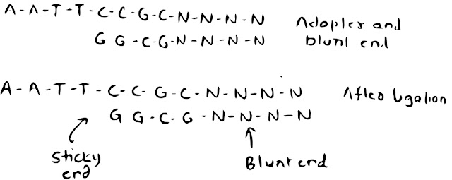
Adaptor: Adaptors are linkers with cohesive ends. 
Blunt end of DNA with blunt end of Adaptors joined by ligase, gives sticky ends. 


Friday, 2 October 2020

Organ of Immune System

Organ of immune system


The parts of body which comprises individual cells, entire organs and organ systems that help in protection from infection of pathogens (foreign bodies).

Picture courtesy: https://clinicalinfo.hiv.gov/en/glossary/immune-system

The organs of immune system are.

    • Skin       
    • Mucous Membrane 
    • Lymphatic System

NOTE: Skin and Mucous Membrane are first line of defence and act as a physical barrier.

Some of Physical barriers are.

  • Antibacterial substance: enzyme present in the Saliva, tears and their ways of respiratory system.
  • Mucus: present in the bronchi that helps to move out germ, bacteria via the hair like projection structure called Cilia.
  • Stomach acid; hydrochloric acid (HCL) present in stomach helps to excrete out or dilute the bacteria and germs.
  • Helping hands bacteria; Bacteria over the skin that helps to maintain the PH and temperature of the body and also kill the other bacteria and organism to protect the body.


Organ of Lymphatic System

  • Primary lymphoid organs
    • Bone marrow
    • Thymus
  • Secondary lymphoid organs
    • Lymph nodes
    • Spleen
    • Tonsils


Primary Lymphoid Organs: 

Differentiation and maturation of lymphocytes takes place.

  • Stem Cell Niche (SCN): The Microenvironment where any stem cells self-generate and differentiate to perform cellular functions very well.

Bone Marrow  

The spongy like a structure in the bone where differentiation and maturation of all Erythroid Cells, Myeloid Cells and B-lymphocytes cells occur.

Friday, 1 May 2020

Circadian Rhythm


      We all make timetable for our work, and we work on that perfect clock time. As the same our body also have a clock known as biological clock and that biological clock produce a circadian rhythm and regulate their timing.


Circadian rhythm:

Circadian rhythm is known as internal clocks that is present within human beings. These internal clocks control our sleep - wake rhythm/cycle. It also controls our metabolism, body temperature, cognition and many more things. There are many factors that regulate and control of circadian rhythm like light, darkness, and aging. Brain has great role in controlling the cycle.
To know more about circadian rhythms

Let's jump little deeper:

In 2017 three scientist name Jeffrey C Hall, Michael Rosbach and Michael young discovered the molecular mechanism of circadian rhythm and they get physiology Nobel Prize 2017.

  These three-scientist discovered that the circadian rhythm is regulated by the period gene which undergoes its oscillation. The PER protein facilitates the oscillation cycle.  These per protein is present in eye tissue, ovaries, brain neurons and in salivary gland. Scientist found that this PER protein work with TIMELESS gene and that gene code for TIM protein. These per protein work as shuttle between cytoplasm and nucleus and that regulates/maintain the level of PER mRNA.


MASTER CLOCK:

Circadian rhythm is related to our brain, and it is called master clock. In human, master clock is a bunch of 20000 neurons that creates a structure called SCN (suprachiasmatic nucleus) these SCN is present in hypothalamus and receives direct light from the eyes.

    Regulation of timing that is circadian rhythm determines our sleeping pattern. Melatonin is a hormone that are responsible for sleep, and the production of melatonin is regulates by SCN that is our master clock.
Circadian rhythm has two clocks, one is central clock that is located in the SCN and other is peripheral clock that is located in all tissue and organ.


Role of light:

When light comes from optic nerve from else to the brain the SCN indicated or stimulates the brain for the production of more melatonin. And thats the reason why people get drowsy. These happen like vice-versa when there is lighter. 


Effect of night shift work: 

Rhythm is disturbed by night shift work and may lead to specific diabetes and cancer. Not only night work but the blue light that comes from your laptop or mobile also disrupt the rhythm by suppressing the production of hormone melatonin by prolong release of PER protein.


Jet lag:

Disruption of circadian rhythm due to traveling to the different time zone is known as jet lag. (scientific term: Circadian dysrhythmia) for jet lag disorder there is no need of any medical treatment because it is a temporary disorder.  Not only light but physical activity also influence of circadian clock. Exercise can improve or reduce the risk of Disruption of circadian rhythmicity. But further research still needs to know the depth of these all reasons.


I Hope you liked it, Thank you :)
@Maulik Ramanandi.

Story of Vanderwaal forces and its help in coagulation.

πŸ§ͺ “The Tale of the Two Tiny Travelers” In the magical world of Colloidia , countless tiny particles—called Colloids —float happily in wate...